
 |
Visiting ProfessorRawil specialized as a visiting scientist in the research group of Prof Paunov at the Department of Chemistry of the University of Hull in 2009 and 2010. He is currently an associate professor at Kazan Federal University.Host: Prof. Vesselin N. Paunov (PI) |
Research project 1:
Building anisotropic cellosomes by templating microcrystals with living cells
We developed anisotropic cellosomes, which are hollow microcapsules whose membranes consist of single monolayer of living cells. These structures were designed based on templating of anisotropic microcrystals of calcium carbonate with living cells, which allowed us to fabricate cellosomes of rhombohedral and needle-like morphologies. Calcium carbonate microcrystals of anisotropic shapes were coated with several consecutive layers of oppositely charged polyelectrolytes to obtain a positive surface charge which was used to immobilise yeast cells coated with anionic polyelectrolyte of their surfaces. After dissolving of sacrificial cores, hollow multicellular structures were obtained. The viability of the cells in the produced structures was confirmed by using fluorescein diacetate. In order to optimize the separation of cellosomes from free cells magnetic nanoparticles were immobilised onto the surface of templates prior to the cells deposition, which greatly facilitated the separation using a permanent magnet. Two alternative approaches were developed to form celloidosome structures using magnetically functionalised core-shell microparticles which resulted in the formation of celloidosomes with needle-like and cubic-like geometries which follows the original morphology of the calcium carbonate microcrystals [1-3].
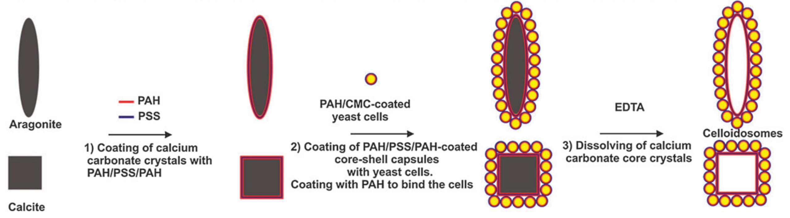
Fig. 1 Schematic representation of experimental approach for the development of cellosomes by templating needle-like (aragonite) and cubic-like (calcite) microcrystals pre-coated with PAH with polyelectrolyte coated cells.

Fig. 2. Optical microscopy images of: (a) – aragonite microcrystal coated with PAH/MNP/PAH/PSS/PAH; (b,c) – needle-like core-shell capsules coated with yeast cells; (d) – needle-like cellosome; (e,f) – rectangular celloidosomes; (g) – calcite microcrystal coated with coated with PAH/MNP/PAH/PSS/PAH; (h-j) – cubic-like core-shell capsules coated with yeast cells; (k,l) – cubic-like celloidosomes.
References
1) Brandy, M.K., Cayre, O.J., Fakhrullin, R. F., Velev, O.D., Paunov, V.N., “Directed assembly of cells into living yeastosomes by microbubble templating”, Soft Matter, 6 (2010) 3494-3498.
2) Fakhrullin, R.F., Brandy, M.K., Cayre, O.J., Velev, O.D., Paunov, V.N., “Live celloidosome structures based on the assembly of individual cells by colloid interactions”, Phys. Chem. Chem. Phys., 12 (2010) 11912-11922.
3) Fakhrullin, R., Paunov, V.N., “Fabrication of living cellosomes of rod-like and rhombohedral morphologies based on magnetically responsive templates”, Chem. Commun, 19 (2009) 2511-2513.
Research project 2:
Making Cyborg Cells: Interfacing Living Cells with Nanoparticles and Polymers
Living cells interfaced with a range of polyelectrolyte coatings, magnetic and noble metal nanoparticles, hard mineral shells and other complex nanomaterials can perform functions often completely different from their original specialization [1]. Such “cyborg cells” are already finding a range of novel applications in areas like whole cell biosensors, bioelectronics, toxicity microscreening, tissue engineering, cell implant protection and bioanalytical chemistry. We developed a direct technique for preparation of magnetically functionalised yeast cells by using polyelectrolyte mediated deposition of magnetite nanoparticles [1,2]. We demonstrate that the cells preserve their viability after the magnetite deposition and show that the magnetic nanoparticles form a multilayered coating on the outer side of the yeast cells wall. We applied our technique to produce magnetically functionalised yeast cells expressing Green Fluorescent Protein (GFP) under the control of RAD54-GFP reporter and demonstrated that their fluorescence emission is not influenced by the presence of magnetite-polyelectrolyte composite coating. We show that the individual cells can be successfully manipulated by an external magnetic field which can be used for their deposition, holding and subsequent removal from microfluidic devices for genotoxicity and cytotoxicity biosensor applications. Our technique for direct magnetization of cells can find many other biotech applications including biosensors, bioreactors and bioseparation [3-4].
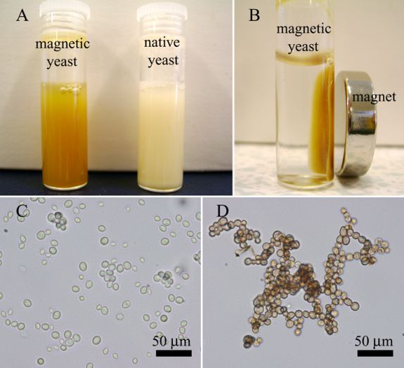
Fig. 1. (A) a photograph of the aqueous suspensions of the native and the magnetic yeast cells; (B) the magnetic behaviour of the magnetized yeast cells illustrated using a permanent magnet; (C) optical microscopy image of the native yeast cells; (D) optical microscopy image of the magnetized yeast cells.

Fig. 2. Transmission electron microscopy images of thin sections of yeast cells coated with PAH/PSS/PAH/TMA-stabilized MNPs/PAH/PSS.
References
- Fakhrullin, R.F., Garcia-Alonso, J., Paunov, V.N., “A Direct technique for preparation of magnetically functionalized living yeast cells”, Soft Matter, 6 (2010) 391-397.
- Garcia-Alonso, J, Fakhrullin, R. F., Paunov, V.N., “Rapid and direct magnetization of GFP-reporter yeast for micro-screening systems”, Biosensors and Bioelectronics, 25 (2010) 1816-1819.
- Zhang, D., Fakhrullin, R.F., Özmen, M., Wang, H., Wang, J., Paunov, V.N., Li, G., Huang, W.E., “Functionalization of whole-cell bacterial reporters with magnetic nanoparticles”, Microbial Biotechnology, 4 (2011) 89-97.
- Fakhrullin, R. F., Zamaleeva, A.I., Minullina, Renata T., Paunov, V.N.,”Cyborg cells: functionalisation of living cells with polymers and nanomaterials”, Chemical Society Reviews, 41 (2012) 4189-4206.
