
 |
Former PhD student of Prof PaunovWill Small graduated as a PhD in 2008 and is currently working as a Lead Applications Scientist at Croda Health Care.PhD Supervisors: Prof. Vesselin N. Paunov (PI) and Marc inhet Panhuis (Co-PI) |
Research project 1 (PhD thesis):
Electrically anisotropic hydrogels with bio-functionalised silver nanowires
We have developed a device based on a microwire network formed from bio-functionalised silver nanowires (AgNWs) through dielectrophoresis (DEP) and hydrogel entrapment. This was achieved by carrying out the DEP assembly of AgNWs in an agarose aqueous solution above its gelling temperature and then cooling to encapsulate the assembled structure within the hydrogel which turns it into an electrically anisotropic material that contains up to 99% water. We have studied in detail the formation of microwires assembled from silver nanowires (AgNWs) in agarose gel, at fixed temperature and AC field voltage, which allowed us to build a “phase diagram” of the microwire assembly as a function of the agarose and AgNW concentration at three different lengths of the AgNWs. In this proof-of-concept study, the AgNWs were functionalised with thiolated biotin, then assembled into microwires by DEP and entrapped into a hydrogel. When exposed to a solution of streptavidin the biosensor device showed a marked increase of the cell conductivity due to compacting of the AgNWs which allows streptavidin detection. This generic type of biosensing device could find applications for detection of other biomolecules, the response to which can be directly converted to an electric signal. Conjugating specific antibodies on the AgNW surface would allow the detection of a matching antigen (e.g. protein) in the solution exposed to the anisotropic hydrogel [1,2].
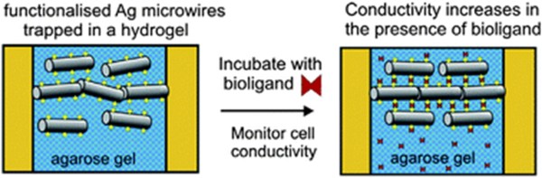
Fig. 1. Optical microscopy images of DEP assembled silver microwires from AgNWs at a concentration of 0.50% w/v in 0.50% w/v agarose gel. (B) DEP assembled microwires from AgNWs of diameter 100 nm. Scale bars represent 1 mm. (C) DEP assembled microwires from AgNWs of diameter 200 nm. Scale bar is 200 mm.

Fig. 2 Plot of the resistance (RHS axis) and the mass (LHS axis) of an agarose gel (0.50% w/v) with encapsulated microwires assembled from 50 nm diameter AgNWs (0.50% w/v) over time. Resistance is measured through the electrodes A– A (filled squares) and B–B (empty squares), while the mass of the film as a percentage of its initial mass is also shown (empty triangles).
References
- Small, W.R., Paunov, V.N., Dielectrophoretic fabrication of electrically anisotropic hydrogels with bio-functionalised silver nanowires, Mater. Chem. B, 1 (2013) 5798 – 5805.
- Small, W.R., Paunov, V.N., “Fabrication of electrically anisotropic agarose gels by dielectrophoretic assembly and encapsulation of silver nanowires”, Mater. Chem., 16 (2008) 2082-2084.
Research project 2:
Scaffold free fabrication of linear multicellular assemblies
We have designed a scaffold-free cell assembly method which can produce linear structures of individual living cells without using templates. The method involves dielectrophoretic assembly of cells suspended in a solution of a gelling agent above the gelling temperature. After the cell assembly in string-like structures was achieved with the AC electric field still applied, we gelled the solution by cooling it below its gelling point. The hydrogel entraps the assembled cell structure, preventing its disassembly and allowing further analysis without the presence of the external electric field. We pre-functionalised the cells with polyelectrolytes before the dielectrophoretic assembly which allowed us to line them up in multicellular strings and bind them together with oppositely charged polyelectrolyte after the gel formation. Finally, by dissolving the hydrogel, we released the linear chains of living cells which were collected and studied by optical and fluorescence microscopy. Cell viability tests with fluorescein diacetate confirmed that the cells in the formed worm-like structures remain viable after the cell assembly procedure. The linear cell aggregates are stable without the electric field and can be further cultured or treated with additional polyelectrolytes which make the method attractive for tissue engineering. We envisage that this technique could find possible applications for assembly of neuron cells in linear structures and more complex cell networks.
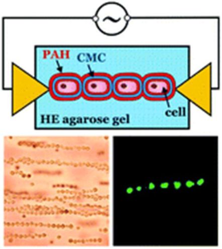
Fig. 1. Schematics of DEP-assisted fabrication of linear cell aggregates and their locking into chains by oppositely charged polyelectrolyte. The LHS image show the cell strings during application of DEP and the RHS fluorescence image shows that the cells retain their viability after the DEP-assisted assembly (FDA assay).
References
- Small, W.R., Paunov, V.N., Scaffold free fabrication of linear multicellular assemblies by dielectrophoretic hydrogel trapping technique, Biomaterials Science, 1 (2013) 996-1002.
